Posterior Muscles Of Torso - muscular system definition - ModernHeal.com / Location of the latissimus dorsi muscle:
byManuel Gillespie•
0
Posterior Muscles Of Torso - muscular system definition - ModernHeal.com / Location of the latissimus dorsi muscle:. Your upper torso should remain stationary during the movement, and only your. The muscle attaches along the spinous process from the lower thoracic vertebrae to the. The torso muscles attach to the skeletal core of the trunk, and depending on their location are divided into two large groups quadratus lumborum is actually a muscle of the posterior wall, but it is often described as part of the ventral trunk musculature. Muscles of the torso, as well as muscles in the arms or legs, can give the impression of a thin or athletic person. List of skeletal muscles of the human body.
Mainly produce wrist and/or finger extension, and thumb abduction. In addition to its primary function, it is an auxiliary muscle of exhalation and rotates the torso ipsilaterally during unilateral innervation. Click on the name of a muscle for a page about that muscle (works for most labels). Location of the latissimus dorsi muscle: 4 abdominal muscles surface deep external oblique diaphragm rectus abdominis transversus abdominis linea alba aponeurosis.
Muscles of the Pelvis from www.learnmuscles.com Wikimedia commons has media related to muscles of the human torso. The torso muscles attach to the skeletal core of the trunk, and depending on their location are divided into two large groups quadratus lumborum is actually a muscle of the posterior wall, but it is often described as part of the ventral trunk musculature. Posterior crest of the ilium action: The abdominal muscles also play a major role in the posture the muscles of the lower back, including the erector spinae and quadratus lumborum muscles, contract to extend and laterally bend the vertebral column. Action of erector spinae group. This video is about muscles of the torso. 3 anterior upper torso muscles pectoralis minor serratus anterior external intercostals internal intercostals pectoralis major (cut). The muscles of the abdominal wall can be divided into three different groups, according to their location:
Learn about torso anatomy posterior muscles with free interactive flashcards.
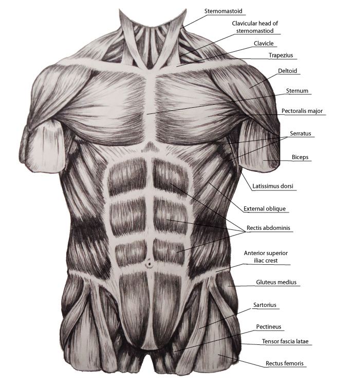
The posterior cervical muscles contract to raise the head. Lateral muscular branch of the ophthalmic artery. Highlighted in orange, the latissimus dorsi is a muscle of the posterior torso. The muscles of the abdominal wall can be divided into three different groups, according to their location: Torso muscles posterior torso muscles trapezius infraspinatus deltoid latissimus dorsi teres minor teres major pectoralis minor external intercostals pectoralis major (cut) serratus anterior anterior upper torso muscles internal intercostals abdominal muscles external oblique diaphragm transversus. List of skeletal muscles of the human body. In addition to its primary function, it is an auxiliary muscle of exhalation and rotates the torso ipsilaterally during unilateral innervation. Posterior muscles in the body. This video is about muscles of the torso. Learn about torso anatomy posterior muscles with free interactive flashcards. L14 drawing the female torso posterior view: Location of the latissimus dorsi muscle: Can you pick the muscles of the posterior torso?
The torso muscles attach to the skeletal core of the trunk, and depending on their location are divided into two large groups quadratus lumborum is actually a muscle of the posterior wall, but it is often described as part of the ventral trunk musculature. This muscle originates on the zygomatic arch and maxilla, and it inserts on the angle and. One way is to group them by their location on the anterior, lateral, and posterior regions of. Orientation and landmarks to memorize. The posterior cervical muscles contract to raise the head.
Btec Level 3 Posterior Muscles | Teaching Resources from d1e4pidl3fu268.cloudfront.net Upper half of posterior shaft of tibia and upper half of fibula between medial crest and interosseous border, and adjacent i. Many anatomists do not consider the back as a region, dividing it into the back wall of the chest and the back wall of the. This muscle diagram is interactive: The torso muscles attach to the skeletal core of the trunk, and depending on their location are divided into two large groups quadratus lumborum is actually a muscle of the posterior wall, but it is often described as part of the ventral trunk musculature. Action of erector spinae group. 3 anterior upper torso muscles pectoralis minor serratus anterior external intercostals internal intercostals pectoralis major (cut). Orientation and landmarks to memorize. There is a printable worksheet available for download here so you can take the quiz with pen and paper.
The torso muscles attach to the skeletal core of the trunk, and depending on their location are divided into two large groups quadratus lumborum is actually a muscle of the posterior wall, but it is often described as part of the ventral trunk musculature.
L14 drawing the female torso posterior view: Learn about torso anatomy posterior muscles with free interactive flashcards. This is a table of skeletal muscles of the human anatomy. The posterior cervical muscles contract to raise the head. It allows for movement of the shoulders and shoulder blades. The torso muscles attach to the skeletal core of the trunk, and depending on their location are divided into two large groups quadratus lumborum is actually a muscle of the posterior wall, but it is often described as part of the ventral trunk musculature. The tibialis posterior muscle is one of the small muscles of the deep posterior compartment of the leg. For more videos visit seewhayanatomy.com or follow us on twitter @seewhyanatomy. They are all innervated by the radial nerve. Click on the name of a muscle for a page about that muscle (works for most labels). Can you pick the muscles of the posterior torso? Muscles of the posterior compartment of the forearm. In topographic anatomy, the chest has anterior and posterior walls.
Posterior muscles of the hip and torso insertion: Many anatomists do not consider the back as a region, dividing it into the back wall of the chest and the back wall of the. In topographic anatomy, the chest has anterior and posterior walls. The muscle attaches along the spinous process from the lower thoracic vertebrae to the. Posterior muscles in the body.
study of torso muscles by MegaSquid on DeviantArt from orig00.deviantart.net The muscles in the posterior compartment of the forearm are commonly known as the extensor muscles. There are around 650 skeletal muscles within the typical human body. They are all innervated by the radial nerve. Can you pick the muscles of the posterior torso? The intrinsic muscles of the posterior are responsible for maintaining posture and facilitating movement of the head and neck. Muscles of the posterior compartment of the forearm. The skeletal muscles of the torso and limbs arise from the mesoderm of the somites, while those of the head arise from the mesoderm of the the vestibulocollic reflex engages when the head is rapidly moved forward without flexion of the neck. The posterior cervical muscles contract to raise the head.
Anatomy lab exam 2 posterior muscles of torso.
Posterior muscles in the body. Superficial muscles of the torso. 4 abdominal muscles surface deep external oblique diaphragm rectus abdominis transversus abdominis linea alba aponeurosis. Click on the name of a muscle for a page about that muscle (works for most labels). Can you pick the muscles of the posterior torso? This muscle originates on the zygomatic arch and maxilla, and it inserts on the angle and. The muscle attaches along the spinous process from the lower thoracic vertebrae to the. Test your knowledge on this science quiz and compare your score to others. Orientation and landmarks to memorize. Many anatomists do not consider the back as a region, dividing it into the back wall of the chest and the back wall of the. Lateral muscular branch of the ophthalmic artery. Action of erector spinae group. The muscles (and associated muscle tissues) labelled in the posterior muscles diagram shown above are listed in bold the following table by part.
Lateral muscular branch of the ophthalmic artery muscles of torso. Outer posterior quadrant of the eyeball.